Saturday, October 31, 2009

Integration and Insulin


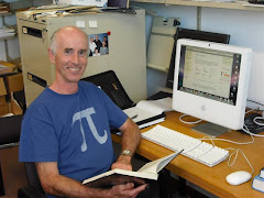
Chris Hann is excited to have used mathematics to play a part in saving lives. At Christchurch Hospital's Intensive Care Unit, a simple practical device developed at Canterbury University's Department of Engineering is being used to help decide how much insulin and food to give patients to stabilise them.


As a result of the high levels of stress, and the action of the hormones produced, critically ill patients often experience hyperglycemia (high blood sugar) and increased insulin resistance. This can lead to further complications, such as organ failure and severe infection. So it is important to control the glucose levels by insulin infusion and a suitable food intake regime.


Various models for glucose-insulin kinetics have been investigated over the years. A model sufficient for this situation can be given by three first order differential equations. G(t) is the concentration of glucose above equilibrium (GE) levels, I(t) is the concentration of insulin (above basal levels) in the blood plasma, and Q(t) is the concentration of interstitial insulin, in the fluid that surrounds the cells - it is this that enables the cells to take up glucose.


The insulin infusion rate u(t) is known.


Notice that the equation for the rate of change of glucose concentration has three terms, the first two giving the decrease in glucose due to insulin (basal plasma insulin and interstitial) and the third giving the increase due to feeding, P(t). The key parameters in the process are the insulin sensitivity, SI, and the glucose fractional clearance, pG, which are patient specific and vary with time. By estimating other less important parameters using typical population values, reformulating the model in terms of integrals and then using numerical integration from the measured data (regular blood glucose levels for patients are taken), a set of equations can be solved for the key parameters over different time periods.


Having worked out a method that is computationally efficient, the model can now be tested with virtual patients, enabling a large number of simulations to be carried out, and refinement of the model. Chris Hann explains that there are more complex models of the glucose-insulin system that could be used, but nothing would be gained from using them in this application, where we are looking at glucose levels on an hourly (or greater) time scale. A detailed and physiologically correct insulin model is only useful for much smaller scales. "We always look to create the simplest possible model for the given application."


Extensive testing showed close agreement between the model predictions and clinical data. The final outcome of the work was the development of the 'Insulin Wheel' and 'Feed Wheel' which give protocols for the administering of insulin and food on the basis of the measured blood glucose levels and the change from the previous hour. Use of this has lead to a significant decline in the mortality rate in the Intensive Care Unit.

Work is now underway on patient-specific modelling of the cardiovascular system in critical care.

My thanks to Dr Chris Hann from Canterbury University. Meeting mathematicians who are enthusiastic about the work they are doing is one of the great pleasures of my fellowship year.

Friday, October 30, 2009

Breast Cancer Detection

The Mechanical Engineering Department at Canterbury University has been developing a new method for detecting breast cancer, which has presented interesting and challenging maths problems. The method has been named DIET - Digital Image-based ElastoTomography.

The rationale for the project is this: Incidence of breast cancer among women in NZ is high, and early detection significantly increases the chances of successful treatment. But the standard current method of breast cancer detection using a mammography machine is not only uncomfortable for women (I have sisters who confirmed that for me!), but involves large and expensive equipment. Screening methods which are less invasive (don’t involve radiation) and equipment which is more portable might increase the up-take of screening.

The concept: Cancerous tissue is stiffer than normal tissue. By subjecting the breast to a rapid fine vibration, it should be possible to detect any region of stiffer tissue from the way the surface moves in response to the vibration.

Five cameras are positioned around the breast to take synchronised images while it is vibrating. (The frequency of vibrations is 20 - 50 Hz and amplitude 1 mm.) Now there is the challenging geometry problem of reconstructing a 3-D image of the breast from the digital images. Once the 3-D motion has been reconstructed comes the problem of using the properties of elastic materials to locate the position and size of any regions of greater stiffness.


Richard Brown, now in the Canterbury University Mathematics and Statistics Dept, was working on this projective geometry problem for his PhD in Engineering. Richard is pictured with Thomas Lotz, who is currently on the team working on the DIET project. Thomas (left) is holding a test silicon 'breast' while Richard (right) is holding a calibration cube, which is used to locate the exact position of the cameras before the test is carried out.


My thanks to Richard and Thomas for introducing me to this work when I was visiting Canterbury University in September.

Sunday, October 25, 2009

Optimisation in Radiotherapy


I first discovered this surprising application of linear programming while looking through annual conference papers of the NZ Operations Research Society (ORSNZ) and have since had the opportunity of talking about the problem with Matthias Ehrgott from Auckland University's Dept of Engineering Science. It is a good example of the way new problems are being presented to mathematicians to work on in this increasingly technological world.


In early external beam radiotherapy treatments, a whole area of the body would be irratiated equally by the beam. Obviously, the maximum radiation dose that could be given to a tumor site has been restricted by the tolerance and sensitivity of the surrounding nearby healthy tissues. An improvement was to create a filter in front of the beam for each patient so as to shape the beam to the tumour. The development of a device called a multileaf collimator (MLC), with moveable 5 mm thick blades (leaves) of lead/tungsten that can block off sections of the beam, together with improvements in imaging techniques, has lead to 3-Dimensional Conformal Radiotherapy, where shaped radiation beams are aimed from several angles of exposure to intersect at the tumour, providing a much larger absorbed dose there than in the surrounding, healthy tissue.

Intensity-Modulated Radiation Therapy (IMRT) is a refinement on conformal radiotherapy, where the intensity beam is controlled, or modulated within the given area, using the MLC. Use of IMRT is growing in more complicated body sites, such as the neck and prostate. Auckland has had this high tech equipment for two or three years.


(Pictured right: multi-leaf collimator)


IMRT in brief:
•Beams (photon/ electron) produced by the linear accelerator are focused on tumour from different directions (3 to 9)
•Intensity across each beam can be modulated by multi-leaf collimator (effectively, beam divided into large no. of beamlets/ bixels)
•Aim:
Focus radiation so that enough dose is delivered to tumour (unlike normal cells, cancerous cells with damaged DNA can’t reproduce) while limiting dose to critical organs and healthy tissue.


The 3 optimisation problems:


1. The geometry problem: What angles (beam directions) should be used?


2. Finding optimal beam intensities for each angle


3. Optimising the delivery schedule


The beam intensity problem can be formulated as a very large linear programme:

Multi-Objective function:
One for tumour, critical organ(s) and normal tissue (minimise underdosing of the tumour and to minimise overdosing healthy organs and other tissues)
Variables:
Let xi be the intensity at bixel i
(the MLC can have 40 leaves and 40 stops, so up to 40 × 40 = 1600 variables for each beam direction)
Constraints:
The region of the body is divided into 3-D volume elements (voxels), and so one constraint for each voxel, given by dose levels of oncologists prescription
(order of 100,000 constraints)
Note: The optimisation computes a set of possible treatment plans for which less overdosing of healthy organs implies more underdosing of the tumour and vice versa. It assists the planner to select one such plan that is best for the patient.

Once the intensities are determined, there is still the problem of how they can be efficiently delivered using the collimator settings, so that the number of shapes and total radiation time is minimised. Effectively, this boils down to the problem of how to decompose a (40 × 40 ) matrix into the sum of matrices whose non-zero elements are identical. No algorithm exists for minimising the number of matrices in the decomposition, so here we have a new problem for computer folks.


The highly complex IMRT treatment has thrown up challenging problems for mathematicians working in OR. Matthias Ehrgott has been working on the multi-objective linear programming problem mentioned above. His time and assistance in giving me an insight into this work has been greatly appreciated.